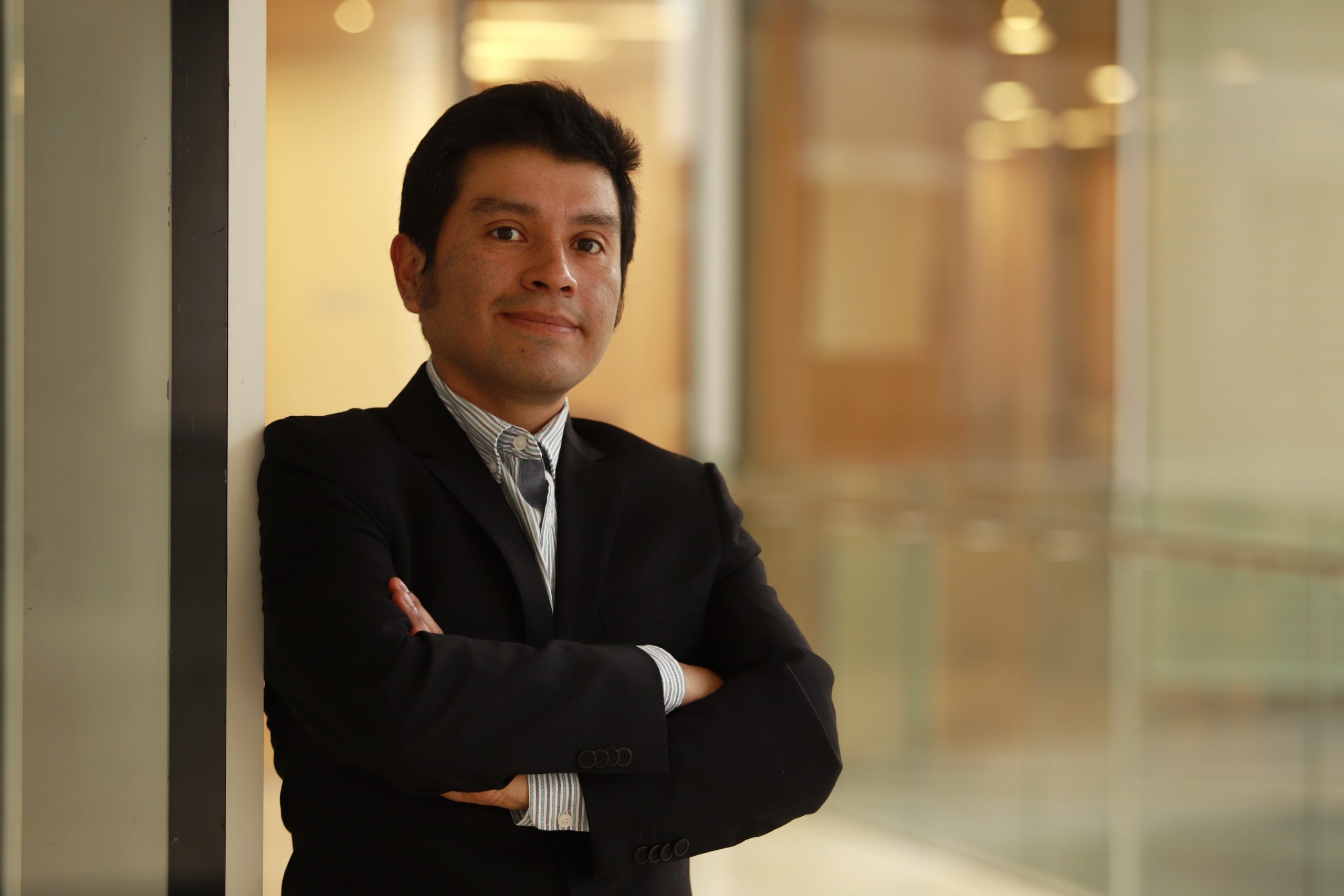
An interdisciplinary team, led by researcher from the School of Engineering and Applied Sciences, José Manuel Saavedra, is developing generative models that allow reconstructing CT images with less than 50% of radiation, without losing diagnostic quality.
A team led by researcher from the School of Engineering and Applied Sciences , José Manuel Saavedra, and radiologist Héctor Henríquez, an academic from the School of Medicine, is developing an innovative AI-based technology: generative models capable of reconstructing computed tomography (CT) images - obtained with significantly reduced radiation doses - while maintaining diagnostic quality comparable to traditional images.
In Chile, CT use has increased by 80% since 2010, according to OECD data. This imaging test combines multiple X-rays, from different angles, to create cross-sectional slices of the body. Thanks to this technology, healthcare professionals can observe organs, bones, blood vessels and soft tissues in great detail, allowing them to detect diseases, internal injuries, tumors and other conditions that require timely medical attention. Its use is essential in emergency contexts, in cancer diagnosis and follow-up, and in complex treatment planning. However, its intensive use also carries a risk: repeated exposure to ionizing radiation has the ability to penetrate the body's tissues and alter the DNA of cells. When exposure is occasional, the body can repair this damage. But when exposure is cumulative - as occurs with frequent examinations or in patients with high clinical follow-up - the risk increases that these alterations will not be adequately corrected, which can lead to cell mutations and, over time, to diseases such as cancer.
"Diffusion generative models are a new class of artificial intelligence algorithms capable of creating detailed and realistic images from simple or incomplete inputs. They work in the inverse way to noise: the model starts from a degraded or noisy image (such as a low-dose CT scan) and, step by step, "cleans it up" until it is transformed into a clear and realistic image, as if it had been captured with a standard dose of radiation. In this project, the model learns to recognize patterns from traditional CT scans and then applies that knowledge to reconstruct high-quality images from versions taken with much less radiation," explains Saavedra, who is also the director of the Computer Vision Laboratory at UANDES.
The academic and researcher adds that reducing radiation dose without losing image quality has been a huge technical and clinical challenge. "Our model has been able to generate accurate images from CT scans taken with more than 50% dose reduction," he says.
Regarding the social impact of this research, Saavedra says: "In addition to improving patient safety, this technology can facilitate access to quality diagnostics in medical centers that do not have state-of-the-art equipment, providing greater equity in the healthcare system."
The project is financed by ANID through the Fondef IDEA program (ID24i10053) and the support of AMIRADI S.A., a national company with more than 30 years of experience in radiology, which contributes with the practical vision of subspecialist radiologists and medical technologists.
The initiative is the result of interdisciplinary work that brings together researchers from different institutions: Violeta Chang; Aline Xavier; Julio Serrano and Anahís Rojas (USACH); José Delpiano (academic and researcher from the School of Engineering and Applied Sciences at UANDES); Tomás de la Sotta (PhD, UANDES) and Camila Figueroa (UCH).

